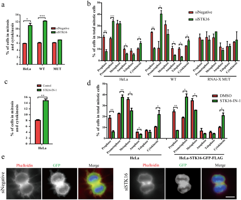
Figure 8. STK16 affects mitotic progression.
(a,b) Quantification of cells in mitosis and cytokinesis, cells in each cell cycle stage during mitosis and cytokinesis in HeLa, HeLa-STK16-GFP-FLAG-WT (WT), and HeLa-STK16-GFP-FLAG-MUT (RNAi resistant mutant, RNAi-X MUT) cells transfected with siSTK16. (c,d) Quantification of cells in mitosis and cytokinesis, cells in each cell cycle stage during mitosis and cytokinesis in HeLa and HeLa-STK16-GFP-FLAG-WT cells treated with 5 μM STK16-IN-1 for 72 hours. (e) Actin spikes and membrane blebbing in cytokinesis cells are reduced after STK16 knockdown. HeLa cells stably expressing STK16-GFP-FLAG-WT were transfected with control or siSTK16 for 3 days before they were fixed and stained by phalloidin and anti-GFP. Experiments were repeated three times and representative images are shown. The data were analyzed using a student’s t- test (*p < 0.05, **p < 0.01, ***p < 0.001). Data show mean ± SEM, n = 3. Scale bar, 10 μm.

