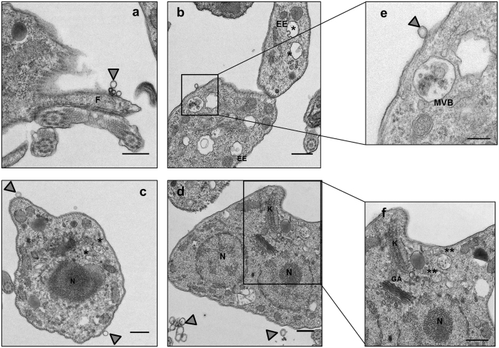
Figure 2. Formation and secretion of EVs in epimastigotes forms.
Formation of multivesicular bodies (MVBs) and secretion of EVs by transmision electron microscopy (TEM). F: fagella; N: nucleus; EE: early endosome; MVB: multivesicular bodies; K: kinetoplastid. **Multivesicular bodies. Grey arrows: Released EVs Size bar = 500 nm.

