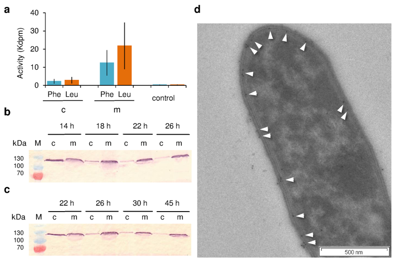
Figure 4. Membrane localization of GS-synthetases in A. migulanus DSM 5759 RC phenotype.
(a) Activity of GrsA and GrsB synthetases in ATP-PPi exchange assays in the membrane (m) and cytoplasmic (c) fractions, which were isolated from cultures grown in G4/4 medium. Phenylalanine and leucine were used as substrates for GrsA and GrsB, respectively. Data are means of three independent experiments ± s.d. (b) Immunodetection of GrsA in the cytoplasmic and membrane fractions of cells grown in G4/4 medium. (c) Immunodetection of GrsA in the cytoplasmic and membrane fractions of cells grown in the minimal GATF1 medium. Data (b,c) are representative of two independent experiments, each. (d) Immunogold staining of GrsA in a GS producing cell from GATF1 medium. White arrows mark the 10 nm gold particles. The figure is representative of at least two independent experiments.

