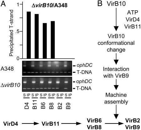
Fig. 5.
TrIP studies with the virB10 strain. (A) TrIP and QTrIP measurements of the T-strand interaction with VirB proteins from the WT or the ΔvirB10 strain. S, supernatant after immunoprecipitation with the anti-VirB antibodies listed on bottom; IP, immunoprecipitated material. For TrIP, the T-strand substrate (T-DNA) and the Ti control fragment (ophDC) were detected by PCR amplification and gel electrophoresis. For QTrIP, quantitative data (Upper) are presented as cpm of incorporated radionucleotide during one cycle in the logarithmic phase of PCR amplication and reported as a ratio of cpm recovered with a given antibody from the virB10 mutant vs. the WT strain. QTrIP data are reported for a single experiment; several repetitions of these experiments showed <5% deviation of the values shown for a given T4SS subunit. (B) Schematic showing the proposed contribution of VirB10 energy coupling to assembly and function of the VirB/D4 T4SS.

