Figure 1.

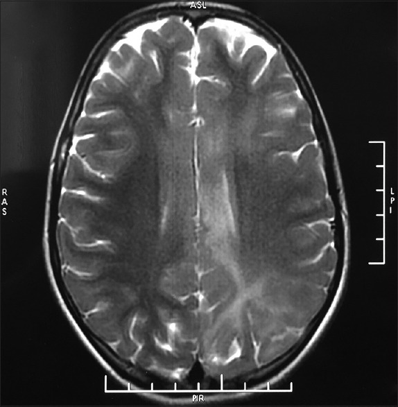
Ill-defined, hyperintense signal changes involving the bilateral fronto–parietal–occipital lobe parenchyma, with loss of the gray–white border distinction in the affected region on T2-weighted images

Ill-defined, hyperintense signal changes involving the bilateral fronto–parietal–occipital lobe parenchyma, with loss of the gray–white border distinction in the affected region on T2-weighted images