
Fig. 3.
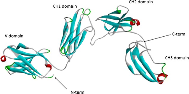
Schematization of the three-dimensional model of sea bass IgT. N- and C-terminus of the model are indicated by arrows. The four structural domains are indicated, from left to right, as VH, CH1-gamma, CH2-gamma, and CH3-gamma, respectively. Short helices are coloured in red, while beta strand are in cyan, and turns in green