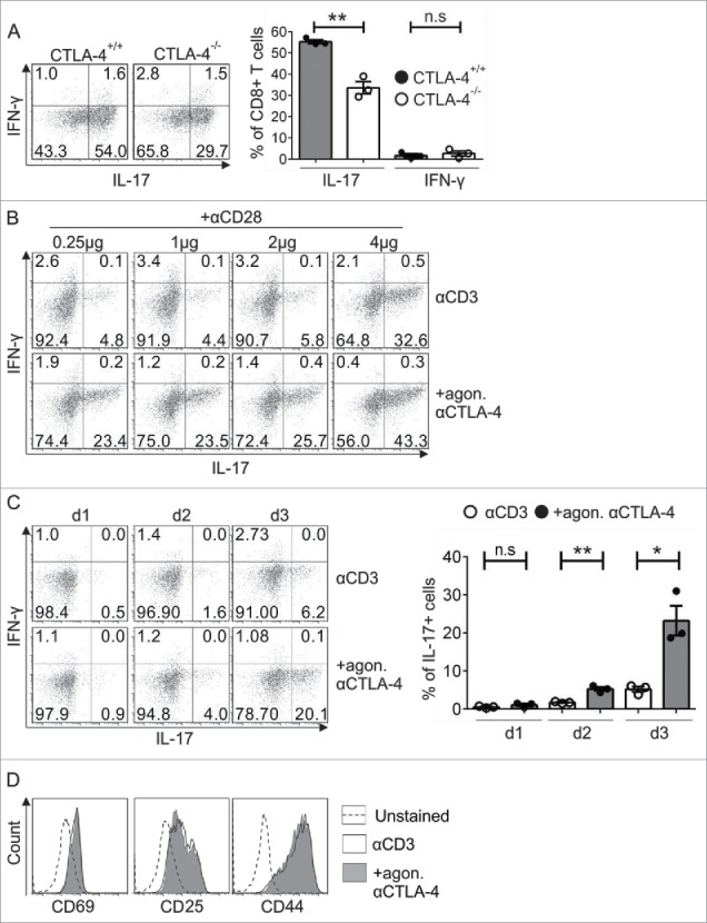
Figure 1.

Analysis of the exclusive role of CTLA-4 in Tc17 differentiation. (A) Naive CD8+ T cells from CTLA-4+/+ and CTLA-4−/− OT.1 mice were activated with the specific antigen OVA257–264 in the presence of APCs under Tc17 conditions. IL-17 and IFNγ expression in these cells was analyzed by flow cytometry for 72 h after primary stimulation (left). Cumulative staining results are shown on the right. The data are representative of three independent experiments. (B) CD8+ T cells from C57BL/6JRj mice were stimulated under Tc17 conditions by crosslinking the cells with plate-bound immobilized anti-CD3 (3 μg/mL) and anti-CD28 (0.25–4 µg/mL) in the presence (+agon. αCTLA-4) or absence (αCD3) of immobilized anti-CTLA-4 (10 μg/mL). Three days after the primary stimulation, IL-17 expression in these cells was analyzed by flow cytometry. The data are from one representative experiment. (C) IL-17 and IFNγ expression in CD3-stimulated (3 μg/mL) cells in the presence or absence of CTLA-4 crosslinking (10 μg/mL) was analyzed by flow cytometry every day until day 3. Cumulative staining results are shown on the right. The data are representative of three independent experiments. (D) CD8+ T cells from C57BL/6JRj mice were cultured as in (C) and analyzed for the surface expression of CD69, CD25 and CD44 on day 3 by flow cytometry. The data are from a single experiment that is representative of three independent experiments. The error bars denote ± SEM. **p < 0.01, *p < 0.05, n.s.: not significant, unpaired t-test.
