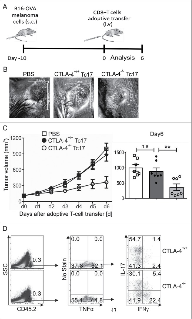
Figure 4.

CTLA-4 regulates the cytotoxic activity of Tc17 cells. (A) Schematic of the tumor experiment. Recipient Ly5.1 mice were s.c. injected with B16-OVA melanoma cells. Approximately 10 d later, when a visible tumor was present, CTLA-4+/+ and CTLA-4−/− OT.1 CD8+ T cells that had been stimulated under Tc17 conditions for 3 d were adoptively transferred into the recipient mice through intravenous (i.v.) injection, and tumor growth was measured for the next 6 d. (B) Pictorial representation of tumor size in the recipient mice on day 6 after adoptive transfer with PBS or CTLA-4+/+ or CTLA-4−/− OT.1 Tc17 cells. (C) Tumor growth in the mice receiving PBS or CTLA-4+/+ or CTLA-4−/− OT.1 Tc17 cells was measured on a daily basis until day 6. Results represent ± SEM of seven mice per group from three independent experiments. Cumulative bar graphs of tumor volume in the recipient mice on day 6 are shown on the right. Results represent ± SEM of seven mice per group from three independent experiments. (D) Adoptively transferred CD45.2+ cells were surface stained ex vivo in the splenocytes of the tumor-bearing mice 6 d after the transfer of CTLA-4+/+ or CTLA-4−/− OT.1 Tc17 cells and were analyzed for TNF-α, IL-17 and IFNγ production by flow cytometry. The data are from one representative experiment. The error bars denote ± SEM. **p < 0.01, n.s.: not significant, Mann–Whitney U-test.
