Figure 1.

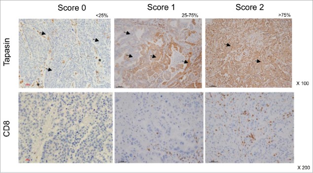
Immunohistochemistry of human primary NSCLC. Representative staining patterns of formalin-fixed, paraffin-embedded tumor lesions with tapasin mAb (upper panels) and CD8+ mAb (lower panels). Tapasin scores and CD8+ scores are determined by percentages of positive tumor cells and by percentages of tumor-infiltrating positive cells in tumor lesions, respectively: 0, <25% positive; 1, 25% to 75% positive; 2, >75% positive. Arrows, tumor cells. Asterisks, stromal (non-tumor) cells. Magnification, x100 and x200.
