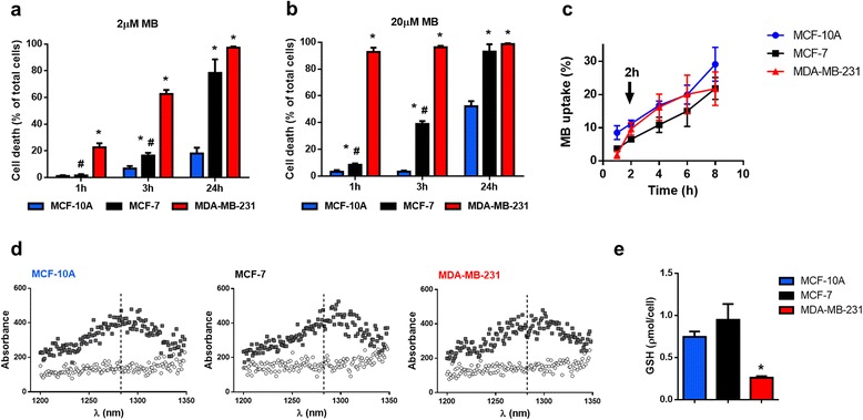
Fig. 1.

MB-PDT induces massive death in tumorigenic cells and weakly affects normal-like cells. Viability time curves after MB-PDT of cell cultures with 2 (a) or 20 μM of MB (b) followed by 4.5 J/cm2 irradiation obtained at 1 h, 3 h and 24 h post-irradiation (n = 3 independent experiments) * p < 0.05 versus MCF-10A; # p < 0.05 versus MDA-MB-231. (c) Curves of MB incorporation in MDA-MB-231, MCF-7 and MCF-10A after 1, 2, 4, 6, and 8 h of incubation (n = 4 independent experiments). (d) Emission spectrum from MB-free MCF-10A, MCF-7 and MDA-MB-231 (white circles); and emission spectra from cells exposed to 20 μM MB for 2 h (gray squares). (e) Cellular GSH levels in MDA-MB-231, MCF-7 and MCF-10A cells * p < 0.05 versus MCF-10A. Results are shown as mean ± s.e.m
