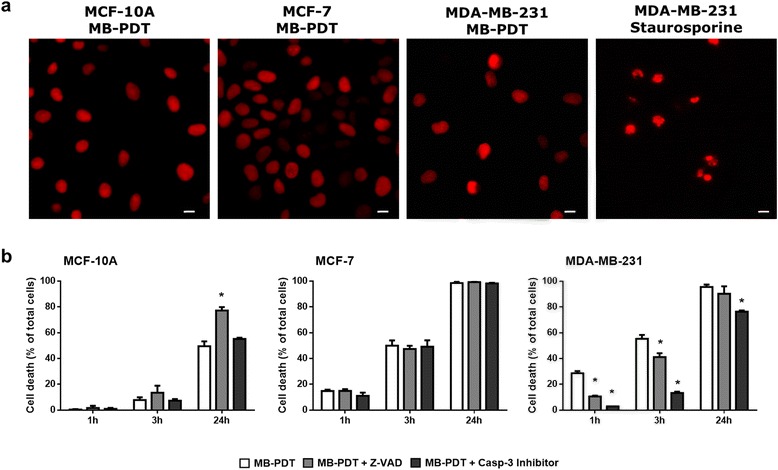
Fig. 2.

Apoptosis pathway is not the main mechanism involved in MB-PDT cell death. (a) Representative image of human mammary cells nuclei treated with MB-PDT or staurosporine (MDA-MB231 cells) stained with propidium iodide. Scale bar: 20 μm (b) Cell viability time curves obtained upon 1 h, 3 h and 24 h post MB-PDT performed in the presence or in the absence of a pan-caspase inhibitor (zVAD) or a caspase-3 specific inhibitor (n = 3 independent experiments) * p < 0.05 versus MB-PDT. Results are shown as mean ± s.e.m
