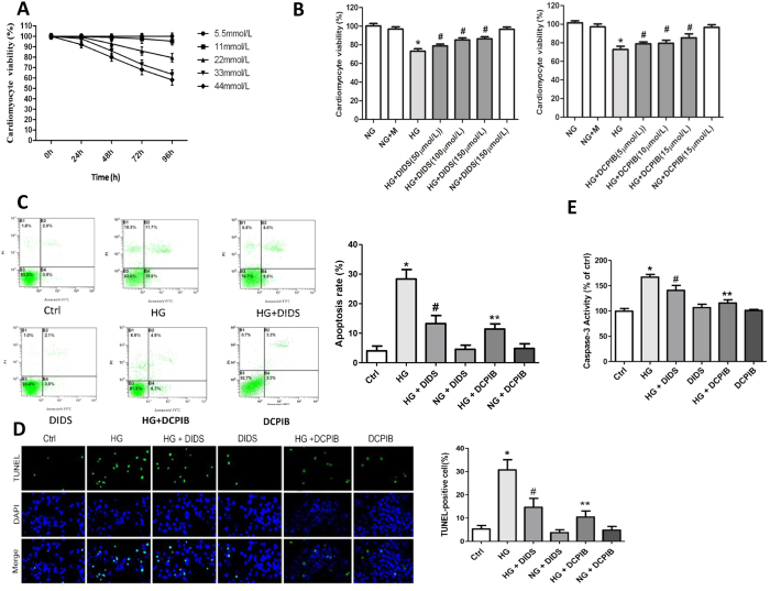
Figure 2. VSOR Cl− channel blockers inhibited cell damage and apoptosis in high glucose-exposed cardiomyocytes.
(A) MTT-assays of cardiomyocytes exposed to indicated concentrations of glucose. n = 12. (B) MTT assays of cardiomyocytes incubated with or without high glucose (HG), DIDS (100 μM) and DCPIB (5 μM). *P < 0.05 v.s. ctrl; #P < 0.05 v.s. HG. n = 5. (C–E) Cardiomyocytes were treated with DIDS (100 μM) or DCPIB (5 μM) for 72 h under the indicated glucose conditions. Cells were subject to Annexin V/PI staining prior to flow cytometry (FCM) analysis (C) and stained for TUNEL. (D) The activities of caspase-3 were measured (E). Data were expressed as mean ± SD and analyzed by two-way ANOVA (n = 4). Scale bar in (D), 200 μm. *P < 0.05 v.s. ctrl; #P < 0.05 v.s. HG. n = 5; **P < 0.05 v.s. HG.

