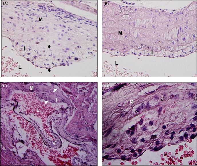
Figure 3.
Atherosclerotic plaque in the H&E stained aortic leaf of sham-infected LDLRnull mice. Panel A is a representative image of plaque in infected LDLRnull mice at 24 weeks of infection at a lower magnification -20× and B is a representative image of plaque in infected LDLRnull mice at a higher magnification 100×. Panel C is a representative image of plaque in sham-infected LDLRnull mice at 24 weeks of infection at a lower magnification 20× and panel D is a representative image of plaque in sham-infected LDLRnull mice at a higher magnification 100×. White arrowheads indicate plaque margin; I indicates intimal layer; M indicates medial layer; V indicates valve; and L indicates lumen.

