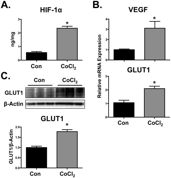
Fig. 1. Activation of HIF-1α signaling in BeWo cells by CoCl2.
Following treatment of BeWo cells with CoCl2 (200 µM) (A) HIF-1α protein levels were quantified by ELISA at 24 h. (B) HIF-1α target gene (VEGF and GLUT1) expression was assessed at 48 h by qPCR and normalized to RPL13A. (C) GLUT1 protein expression was determined by western blot and β-ACTIN was used as a loading control. Data are presented as mean ± SE (n=3-4). Asterisks (*) represent statistically significant differences (p<0.05) compared to control cells (Con).

