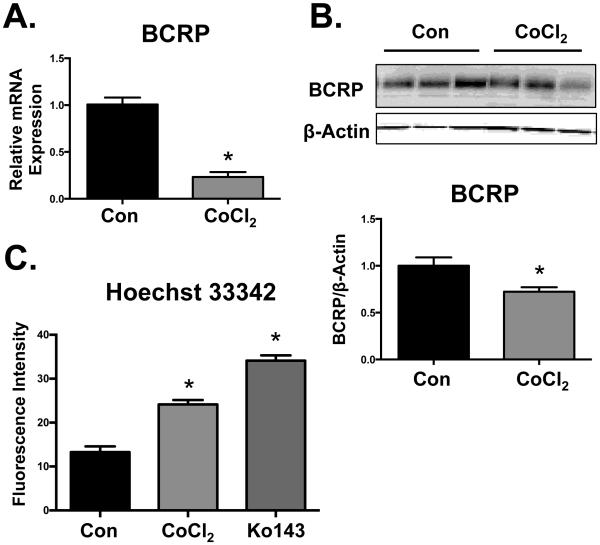
Fig. 2. Down-regulation of BCRP in BeWo cells by CoCl2.
BeWo cells were treated with CoCl2 (200 µM) for 48 h, following which they were processed for (A) BCRP and RPL13A mRNA expression by qPCR. (B) Protein expression of BCRP was determined by western blot and β-ACTIN was used as a loading control. (C) BCRP function was assessed by measuring the cellular retention of Hoechst 33342 (5 µM) in the presence or absence of the BCRP-specific inhibitor (1 μM Ko143). Intracellular fluorescence was quantified by a Cellometer Vision automated cell counter. Data are presented as mean ± SE (n=3-4). Asterisks (*) represent statistically significant differences (p< 0.05) compared to control cells (Con).

