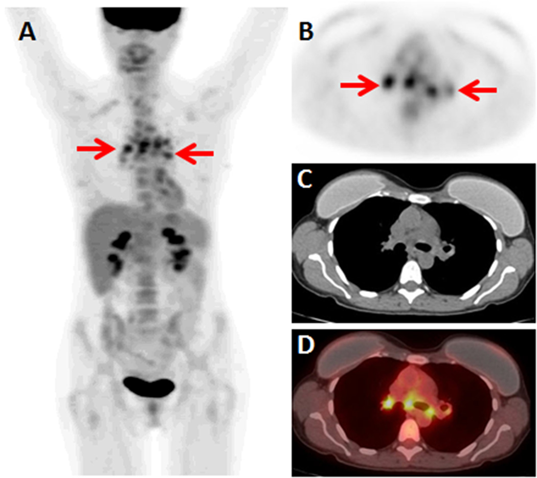
Figure 1.
48-year-old woman with invasive ductal breast cancer. FDG PET/CT was ordered for systemic staging. (A) FDG PET MIP demonstrates multiple FDG-avid foci in the medial thorax (arrows). (B) Axial FDG PET, (C) axial non-contrast CT, and (D) axial FDG PET/CT demonstrate the FDG-avid foci localize symmetrically to the mediastinum and bilateral hila (arrows), without corresponding masses on CT. These findings were called nodal metastases on the initial report. Second opinion report called these findings likely benign, noting that bilateral hilar and mediastinal nodal metastases without axillary or internal mammary nodal metastases would be highly unlikely. A mediastinal biopsy was performed, yielding a diagnosis of sarcoidosis.

