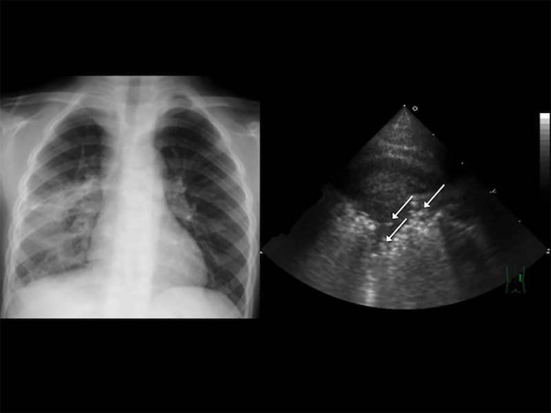
Fig 2. A 4-year-old boy with a high fever and dyspnea, classified as having severe pneumonia in our study.

(A) Chest radiography revealed right lower lobe consolidation. (B) Transthoracic ultrasonography showed multiple hyperechoic spots in the right anterior lower lung, indicating the presence of air bronchograms (arrows).
