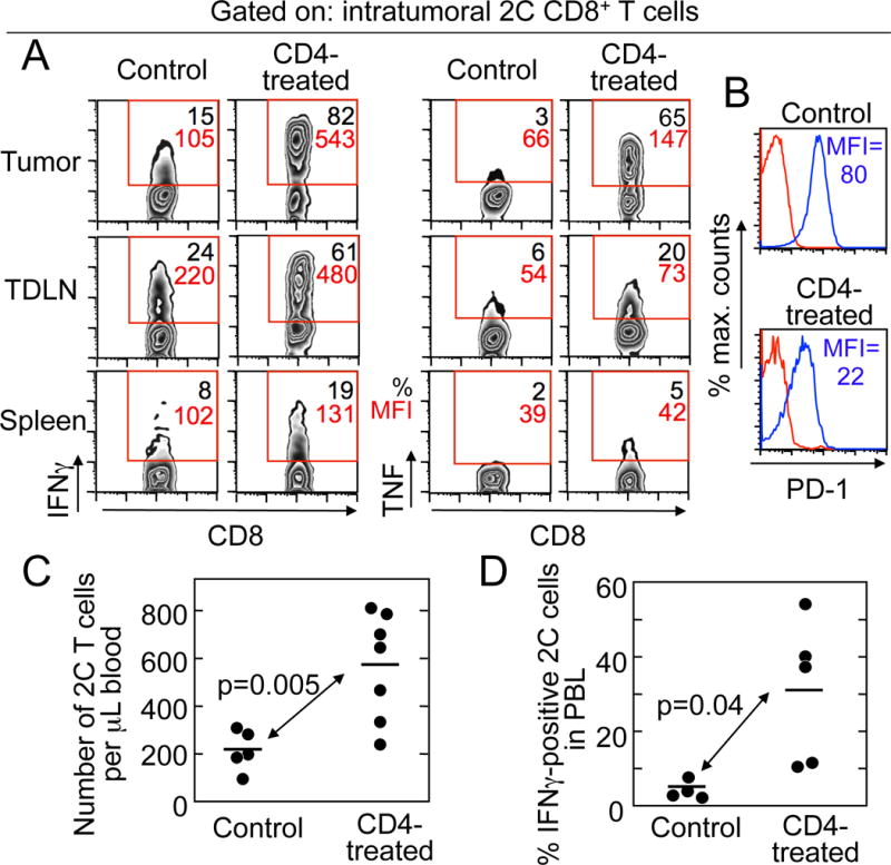
Figure 5. CD4+ T cells restored the function of exhausted CD8+ T cells.

A. CD4+ T cells increased IFNγ and TNF production by tumor-infiltrating CD8+ T cells. IFNγ and TNF were measured in 2C cells recovered from lymphoid organs and PRO4L-SIY-EGFP tumors from OT1-Rag−/− mice with tumors in equilibrium treated or not with allogeneic CD4+ T cells (day 24 after CD4+ T-cell transfer). For each plot the percentage of 2C cells (CD8+Vb8+) producing cytokines is shown in black and the mean fluorescence intensity (MFI) in red. B. CD4+ T-cell transfer reduced the amount of PD-1 on 2C cells (CD8+Vb8+) from tumors. Red: Isotype control, Blue: anti-PD-1. C. CD4+ T cells increased the absolute numbers of 2C CD8+ T cells in the peripheral blood of mice bearing PRO4L-SIY-EGFP tumors. D. CD4+ T-cell treatment increased the production of IFNγ by circulating 2C cells. Mice received CD4+ T cells at days 29–76 after receiving 2C cells, and were bled 13–49 days after CD4+ T-cell transfer. For (A) and (B), 1/3 experiments with consistent results are shown. Data shown in (C) are from 3 independent experiments and 12 mice, and in (D) from 3 independent experiments and 9 mice.
