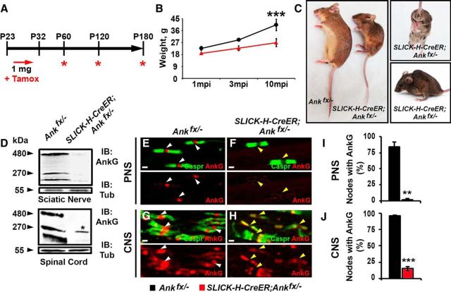
Figure 3.
Juvenile neuron-specific AnkG ablation model. A, Schematic representation of tamoxifen injections for AnkG ablation from adult myelinated axons. Tamoxifen was injected starting at P23 for 10 consecutive days to SLICK-H-CreER;Ankfx/− and Ankfx/− control mice, which were analyzed 1, 3, and 10 mpi. B, Graph representing the weight of Ankfx/− and SLICK-H-CreER;Ankfx/− mice (n = 6 mice/genotype; two-way ANOVA, Bonferroni's post hoc analysis). C, Photographic depiction of 8 mpi SLICK-H-CreER;Ankfx/− mutant and Ankfx/− control mice after AnkG ablation by tamoxifen. D, Immunoblot analysis of SN and SC lysates from 1 mpi Ankfx/− and SLICK-H-CreER;Ankfx/− mice with antibodies against AnkG and α-tubulin (Tub). Asterisk in the SLICK-H-CreER;Ankfx/− lane indicates a 270 kDa AnkG isoform expressed by oligodendrocytes. E–H, Immunostaining of 1 mpi teased SN fibers (E, F) and SCs (G, H) with antibodies against Caspr (green) and AnkG (red) from Ankfx/− and SLICK-H-CreER;Ankfx/− mice. White and yellow arrowheads indicate AnkG-positive and AnkG-negative nodes, respectively. I, J, Quantification of the percentage of nodes with AnkG in 1 mpi Ankfx/− and SLICK-H-CreER;Ankfx/− SNs and SCs, respectively (n = 3 mice/genotype; 120–130 nodes/animal; unpaired, two-tailed Student's t test). Scale bar, 2 μm. All data are represented as the mean ± SEM. *p < 0.05; **p < 0.01; ***p < 0.001.

