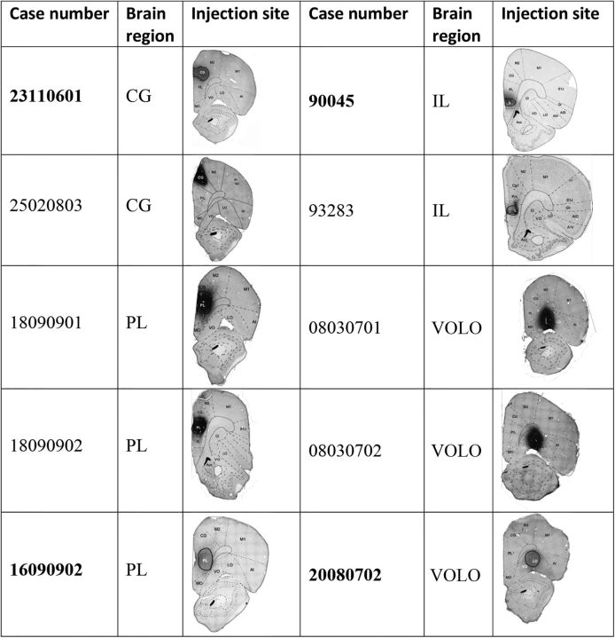
Figure 1.
List of animal cases and injection sites. Selected cases for individual illustration are shown in bold. Photomicrographs of coronal sections are centered on the maximal extent of the tracer. The delineation of the structures was made using the atlas of Paxinos and Watson (1986) as a reference. AI (V, D), agranular insular cortex (ventral and dorsal); Cl, claustrum; GI, granular insular cortex; M1 and M2, motor cortex 1 and 2; MO, medial orbital cortex; primary somatosensory cortex, jaw region.

