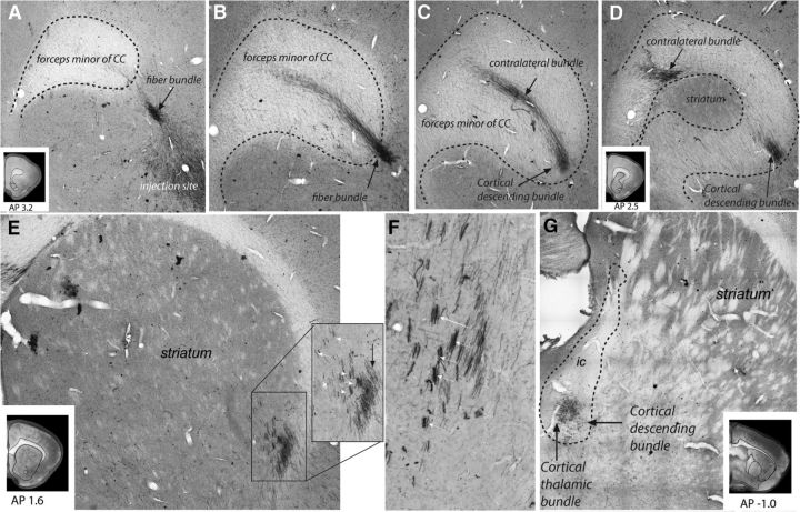
Figure 3.
Photomicrographs of OCIP descending pathways. A–D, Photomicrographs of coronal rat brain sections centered on the right CC at different anteroposterior levels with A the most rostral and D the most caudal section. OCIP (for this illustration, an LO case) axons leave the injection sites and travel toward the CC, where they split into a contralateral and a descending bundle. E, F, OCIP fibers enter the striatum (black arrow) and divide into small fascicles (white arrows) to travel toward the internal capsule. G, OCIP axons embedded within the internal capsule. At this level, two separate bundles can be distinguished: a dense cortical thalamic bundle and a cortical bundle that will continue to descend. AP, Anterior–posterior distance from bregma (mm).

