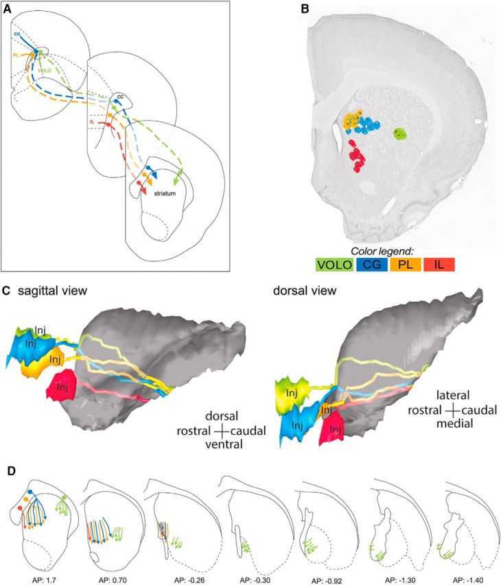
Figure 8.

Topographical organization of OCIP descending pathways. A, Schematic of coronal sections illustrating topography of OCIP bundles within the CC/subcortical WM from the injection site (top left section) to the location where they enter the striatum (bottom right section). Note that the four bundles remain segregated and topographically organized according to the position of structures within PFC. B, Coronal section showing the organization of the labeled OCIP fascicles in the rostral striatum. OCIP bundles descending to the thalamus and brainstem are well segregated within this structure except for CG and PL, which are partly intermixed. C, Sagittal (left) and dorsal (right) 3D views of the striatum showing the topography of OCIP pathways through this structure. D, Schematics of coronal sections illustrating OCIP bundle organization from the location where they enter the striatum to where they enter the internal capsule. Note that the four bundles remain segregated and topographically organized according to the position of structures within PFC, with IL, PL, and CG medial and VOLO lateral. AP, Anterior–posterior distance from bregma; Inj, injection site.
