
Figure 1.
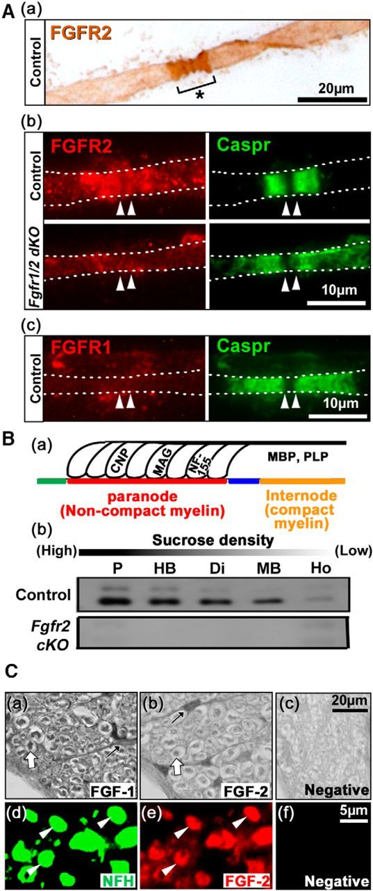
FGFR2 expression is enriched at the paranodal loops of myelin, and FGF1 and FGF2 are expressed by the axons. Aa, Immunolabeling of teased fiber preparations from spinal cords of 2-month-old control mice shows intense staining for FGFR2 in a pattern indicative of its enrichment at the paranodal region (asterisk) of the myelinated fiber. Ab, Double immunolabeling shows that FGFR2 (red) is largely colocalized with the paranodal marker Caspr (green) and that its signal is lost in the Fgfr1/Fgfr2 dKO mice, demonstrating the specificity of this staining pattern for myelin (arrowheads mark the nodal region). Ac, Double immunolabeling shows that anti-FGFR1 or the secondary antibody alone (red) does not label the Caspr+ (green) paranodal region of control mice. Ba, Schematic representation of subdomains of the myelinated fiber [node (green), paranode (red), juxtaparanode (blue), and internode (orange)] and example of the myelin proteins expressed in noncompact (CNP, MAG, NF-155) and compact myelin (MBP, PLP) that distribute in a typical pattern of relative enrichment when myelin is fractionated on a sucrose density gradient (adapted from Menon et al., 2003). Bb, Immunoblotting of fractionated myelin from normal control mice show that FGFR2 is most enriched at high and least enriched at low sucrose densities in a pattern of distribution typical for other noncompact myelin proteins, while FGFR2 is completely lost from all fractions of myelin in the Fgfr2 cKO mice. Ho, Homogenate; Di, dispersion; P, pellet. Ca–Cf, Immunolabeling of spinal cord cross-sections from control mice with anti-FGF1 (a) and anti-FGF2 (b) shows darkly stained regions surrounded by unstained ring-like region (white arrows) in the white matter. Anti-FGF1 and anti-FGF2 also immunolabel astrocyte-like cells (black arrows). Cervical spinal cord sections double-labeled for the axonal marker NFH (d, green) and anti-FGF2 (e, red) show overlapping labeling of axons (arrowheads). Negative controls are secondary antibodies alone (c, f). Representative images taken from similar regions of lateral-ventral white matter are shown. N = 3–4 mice for each condition.