
Figure 10.
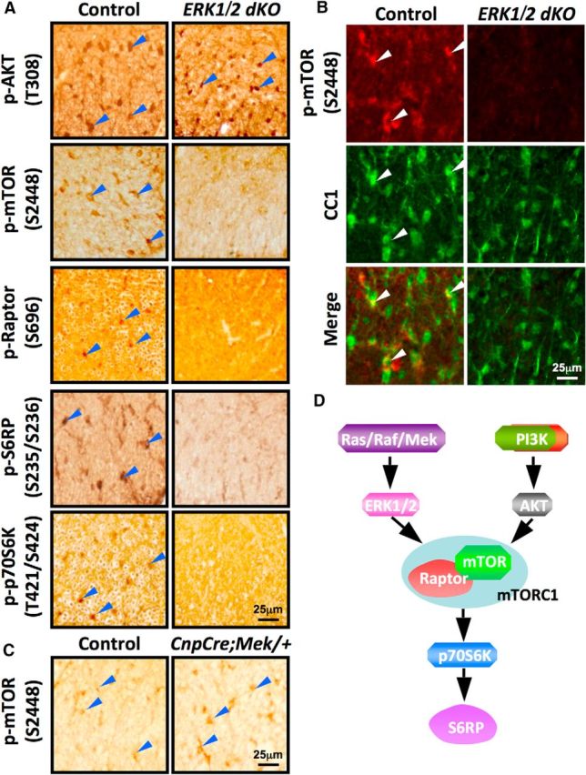
Downregulation of p-mTOR, p-Raptor, p-p70S6K, and p-S6RP but not p-Akt in mice lacking ERK1/2. A, Cervical spinal cord sections from P30 control and ERK1/2 dKO mice analyzed by immunolabeling for p-AktT308, p-mTORS2448, p-RaptorS696, p-S6RPS235/S236, and p-p70S6KT421/S424 show that while the intensity of cellular staining of AktT308 remains unchanged, the signals of p-mTORS2448, p-RaptorS696, p-S6RPS235/S236, and p-p70S6KT421/S424 are decreased dramatically in the white matter of ERK1/2 dKO mice compared to control mice. Blue arrowheads point to cellular staining in the spinal cord white matter. B, Double immunolabeling of cervical spinal cord sections from control and ERK1/2 dKO mice show presence of p-mTORS2448 signal colocalized with CC1 in controls (white arrowheads) and its virtual absence in CCI+ oligodendrocytes in the white matter of the ERK1/2 dKO mice. C, Cervical spinal cord sections from control and CnpCre;Mek/+ mice immunolabeled for p-mTORS2448 at P30 show cellular staining in the control mice and an increase in the intensity of the signal in CnpCre;Mek/+ mice compared to controls. Representative images from the analysis of at least 3 animals per genotype taken from similar regions of lateral-ventral white matter are shown. Blue arrowheads point to cellular staining in the spinal cord white matter. D, Schematic representation of our working model showing convergence between the Mek/ERK1/2–MAPK and the P13K/Akt/mTOR pathways at the level of mTORC1 in mature oligodendrocytes during developmental myelination.