Figure 3.
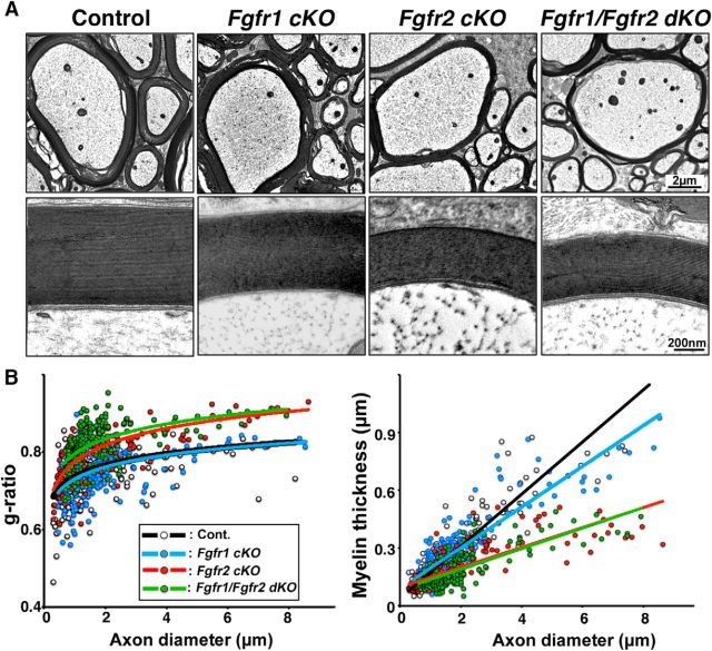
Myelin thickness is reduced in mice lacking Fgfr2 but not Fgfr1. A, Lower- and higher-magnification EM images taken from similar regions of ventral spinal cords at 5–7 months from control, Fgfr1 cKO, Fgfr2 cKO, and Fgfr1/Fgfr2 dKO mice show that while the thickness of myelin is similar in the control and Fgfr1 cKO mice, axons are wrapped by thinner myelin sheaths in Fgfr2 cKO and Fgfr1/Fgfr2 dKO mice. B, As a measure of myelin thickness, scatter plots of g-ratios (left) and myelin thickness (in micrometers; right) are shown for individual fibers in relation to respective axon diameters, confirming that Fgfr2 cKO and Fgfr1/Fgfr2 dKO mice have thinner myelin (higher g-ratios) than Fgfr1 cKO mice and controls. Approximately 200 axons from two mice of each genotype were analyzed from similar regions of the ventral white matter.

