
Figure 4.
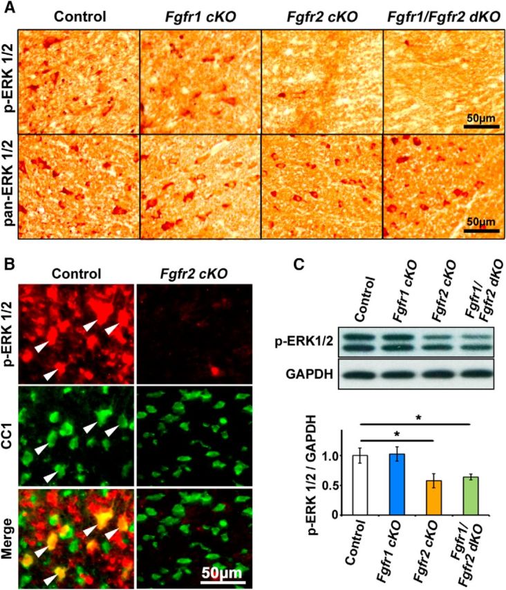
ERK1/2 activity is downregulated in oligodendrocytes in mice lacking Fgfr2 but not Fgfr1. A, Transverse sections of cervical spinal cords at P15, immunolabeled for p-ERK1/2 or pan-ERK1/2, show strong oligodendrocyte-like cellular staining in the white matter of control and Fgfr1 cKO mice, but it is downregulated in Fgfr2 cKO and Fgfr1/Fgfr2 dKO mice. Parallel sections of spinal cords immunolabeled for pan-ERK1/2 show positive signal in all genotypes. B, Double immunolabeling of cervical spinal cord sections from control and Fgfr2 cKO mice show presence of p-ERK1/2 signal colocalized with CC1 in controls (arrowheads) and its virtual absence in CCI+ oligodendrocytes in the matched regions of lateral-ventral white matter of the Fgfr2 cKO mice. C, Immunoblotting of equal amounts of total proteins from homogenates of white matter from spinal cords and quantification of the band intensity on the blots show a statistically significant reduction of p-ERK1/2 levels in Fgfr2 cKO and Fgfr1/Fgfr2 dKO but not Fgfr1 cKO mice compared to control mice. GAPDH, used as a loading control, does not show a change. Three mice from each genotype were analyzed. Error bars represent SEM (N = 3). *p < 0.05.