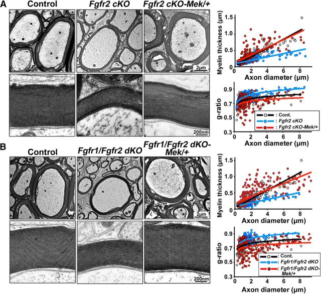
Figure 6.
Reduction of myelin sheath thickness in mice lacking Fgfr2 is rescued by the elevation of ERK1/2 activity in Fgfr2-deficient oligodendrocytes. A, EM images taken from similar ventral regions of cervical spinal cords at low and high magnification at 5 months show reduction of myelin thickness in Fgfr2 cKO mice compared to controls and an increase in Fgfr2-KO;Mek/+ mice. As a measure of myelin thickness, scatter plots of g-ratios and myelin thickness (in micrometers) are shown in relation to axon diameters, confirming the rescue of myelin thickness in Fgfr2-KO;Mek/+ mice. B, Low- and high-magnification EM images and quantification show a reduction of myelin thickness in Fgfr1/Fgfr2 dKO mice and an increase in Fgfr1/Fgfr2-dKO;Mek/+ mice compared to controls. Approximately 200 axons from 2 mice of each genotype were analyzed from similar regions of the ventral white matter.

