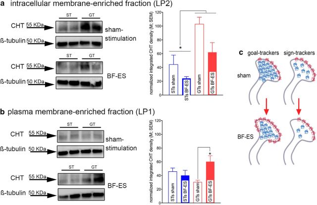
Figure 2.
Effects of BF-ES on subcellular CHT distribution in cortical synaptosomes (n = 19; n = 5 GTs and 5 STs for BF-ES; n = 4 GTs and 5 STs for sham stimulation; the immunoblots depict duplicates from 1 ST and 1 GT). a, Consistent with previous research, at baseline (sham stimulation), in GTs and STs, the majority of CHTs was located on intracellular domains, such as vesicular and endosomal membranes. BF-ES lowered the intracellular density of CHTs in both phenotypes [main effect, no interaction with phenotype; sham, 65.28 ± 12.82 (normalized density); BF-ES, 45.77 ± 9.86]. Moreover, STs generally exhibited lower levels of intracellular CHTs than GTs (main effect of phenotype). b, Whereas BF-ES increased the density of CHTs in the plasma membrane-enriched LP1 fraction in GTs, this was not the case in STs. Results from additional analyses are illustrated in c. In synaptosomes from sham-stimulated animals, the LP2/LP1 ratio was already near 1 (indicated by red protein symbols in membrane). Previous findings indicated that a ratio of 1 represents the limit of CHT mobilization to plasma membrane. Thus, synaptosomal CHT distribution in STs may have already reached this maximum at baseline, primarily attributable to a lower intracellular CHT density than in GTs. Second, in STs, the BF-ES-induced loss of LP2 CHTs (blue protein symbols) was not reciprocated with an increase in LP1 CHTs, suggesting stimulation-induced translocation of a portion of intracellular CHTs to domains other than synaptic plasma membrane and those captured in the present LP2 fraction.

