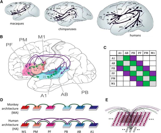
Figure 1.
A, Illustration of perisylvian connectivity structure in macaques, chimpanzees, and humans as revealed by tractography studies [adapted by permission from Macmillan Publishers Ltd: Nature Neuroscience (Rilling et al., 2008), copyright 2008]. Note the strong frontotemporal connectivity of the latter, especially through the dorsal AF curving around the sylvian fissure, and the presence of ventral connections in both. B, A human brain schematic is used to illustrate the area subdivision of the primate frontotemporal perisylvian cortex into M1, PM, and PF, and A1, AB, and PB areas (Garagnani et al., 2008). Green arrows give the connections available in both the human and monkey architecture (HA, MA); purple arrows give connections unique to the human architecture. The purple arrows present only in the HA are meant to reflect the additional connectivity strength available only in humans, as shown by comparative DTI/DWI studies (see main text for detailed discussion). C, Connectivity matrix schematizing the connections according to next-neighbor (green) and indirect, jumping links (purple) skipping one intermediate area. D, Schematic depiction of the neural network architectures, equivalent to B. E, Microstructure of the connectivity of one single excitatory cell (labeled “e”). Local (lateral) inhibition is implemented by an underlying cell “i” (representing a cluster of inhibitory interneurons situated within the same cortical column), which receives excitatory input from all cells situated within a local (5 × 5) neighborhood (dark-colored area) and projects back to e, inhibiting it. Within-area sparse excitatory links (in gray) to and from e are limited to a (19 × 19) neighborhood (light-colored area); between-area excitatory projections (green and purple arcs) are topographic and target 19 × 19 neighborhoods in other areas (not depicted). Panel B has been adapted from Garagnani and Pulvermüller (2013); panels D and E have been adapted from Cortex, 57, Pulvermüller, F. and Garagnani, M., “From sensorimotor learning to memory cells in prefrontal and temporal association cortex: A neurocomputational study of disembodiment”, pp. 1–21, copyright 2014, with permission from Elsevier.

