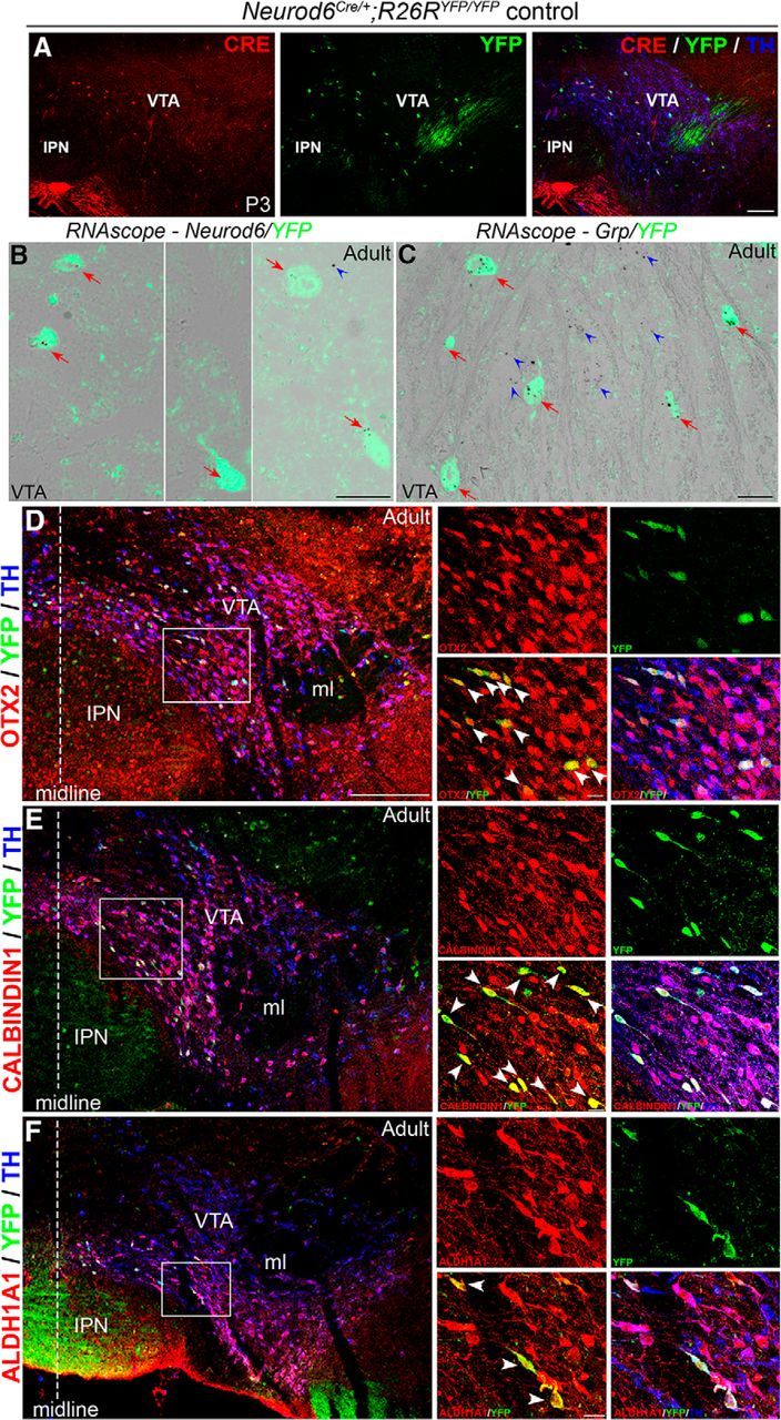
Figure 2.

Identification of a novel subset of mDA neurons in the VTA that expresses Neurod6, OTX2, CALBINDIN1, ALDH1A1, and Grp. A, Double antibody labeling showing that CRE is coexpressed with YFP in TH+ mDA neurons in the VTA of Neurod6Cre/+;R26RYFP/YFP pups at P3. B, In situ hybridization of Neurod6 combined with immunohistochemistry for YFP showing that all YFP+ cells express Neurod6 transcripts (red arrows) in adult Neurod6Cre/+;R26RYFP/YFP mice. A few YFP− cells also express Neurod6 transcripts (blue arrowheads). C, In situ hybridization of Grp combined with immunohistochemistry for YFP showing that Grp transcripts are detected in both YFP+ (red arrows) and YFP− (blue arrowheads) cells in the VTA of adult Neurod6Cre/+ mice. D–F, Triple antibody labeling showing that all YFP+/TH+ mDA neurons express OTX2 (D), CALBINDIN1 (E), and ALDH1A1 (F) in the VTA of adult Neurod6Cre/+;R26RYFP/YFP mice. IPN, Interpeduncular nucleus; ml, medial lemniscus. Dotted vertical lines indicate the midline of the section. White arrowheads indicate triple-labeled cells observed in the red and green channels only. Scale bars: A, 100 μm; B, C, 200 μm (higher magnifications in D–F); D–F, 20 μm.
