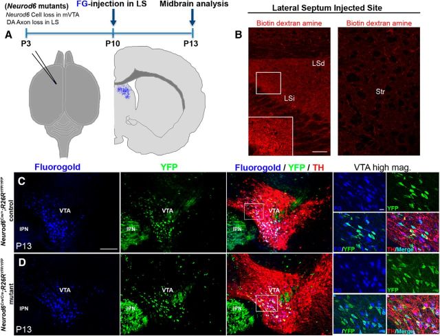
Figure 6.
FG retrograde labeling experiments show that Neurod6+ mDA neurons project to the dorsal and intermediate region of the lateral septum. A, Schematic diagram indicating the position of FG injection in the injected brain and the schedule of the experiment. B, BDA is detected specifically in the LSi as well as the LSd but not in the adjacent striatal regions. C, D, Injection of FG into the septal region results in its retrograde transport of FG into YFP+/TH+ mDA cell bodies in Neurod6 control and mutant mice by P13. Small panels indicate higher magnification of the corresponding boxed regions. IPN, interpeduncular nucleus; mag, magnification. Scale bars: B, 100 μm; C, 200 μm (20 μm, higher magnification).

