Figure 7.
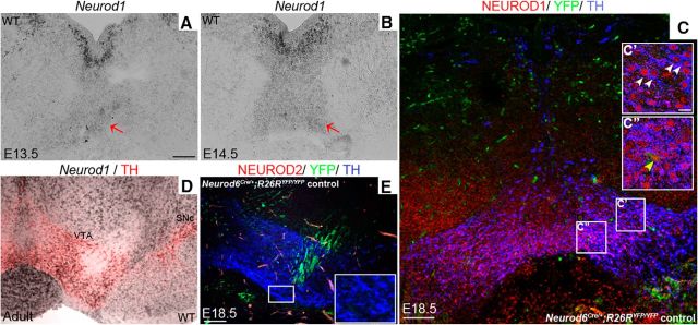
Neurod1, but not Neurod2, is expressed in mDA neurons. A, B, In situ hybridization shows that Neurod1 transcripts are expressed strongly and weakly, respectively, in immature and mature (red arrows) in mDA neurons located ventral to the floor plate of the midbrain at E13.5 (A) and E14.5 (B). C–E, This expression, detected by triple immunolabeling with NEUROD1, TH, and YFP antibodies, is maintained in mature YFP+ and TH+ mDA neurons at E18.5 (C–C″) and in adult (D) Neurod6 control mice by in situ hybridization of Neurod1 followed by immunohistochemistry of GFP and TH. E, In contrast, NEUROD2 was not detected in mDA neurons at E18.5 by immunohistochemistry in sections of central mDA regions. C′, C″, D, Insets, Higher magnification of corresponding boxed areas. C′, White arrowheads indicate double-labeled cells. C″, Yellow arrowhead indicates triple-labeled cells. Scale bars: A, D, 100 μm; C′, 10 μm; E, 100 μm.

