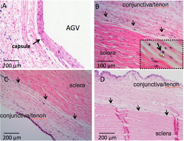
Fig 5. Histological study of tissue reaction to the microfluidic meshwork in comparison with AGV 3 months post implantation.
A. capsule beneath the plate of AGV; B. minimal reaction to the meshwork in rabbit 1, inset figure is a magnified view to a single channel of the meshwork (400x); C. minimal reaction to the meshwork in rabbit 2; D. minimal reaction to the meshwork in rabbit 3. Arrows in B, C and D is to delineate the meshwork.

