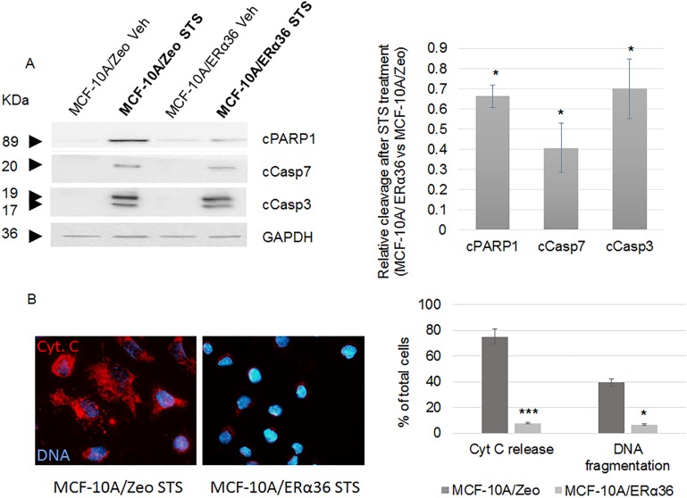
Fig 3. ERα36 overexpression stimulates apoptosis resistance.
MCF-10A/Zeo and MCF-10A/ERα36 cells were exposed to 0.25μM staurosporin (STS) or vehicle (Veh) for 6 hours. A. Cleavage of PARP1 (cPARP1), Caspase 7 (cCasp7) and Caspase 3 (cCasp3) were evaluated with specific antibodies (left panel). GAPDH was used as a loading control. Results depicted in the corresponding histogram are represented as STS versus Vehicle ratio (right panel). ERα36 overexpression triggered a significant 34%, 60% and 30% decrease of PARP1, Caspase 7 and Caspase 3 cleavage, respectively. Each bar represents mean ± S.E.M. N = 4. *: P <0.05. B. Cytochrome c (Cyt. C) release (red, AlexaFluor 555) and DNA fragmentation were respectively assessed by immunofluorescence and TUNEL assay after STS exposure in MCF-10A/Zeo and MCF-10A/ERα36 cells (left panel), then quantified as shown in the corresponding histogram (right panel). No cytochrome c release or DNA fragmentation can be detected in untreated cells (not shown). Each bar represents mean ± S.E.M. N = 3. *: P <0.05, ***: P <0.001.

