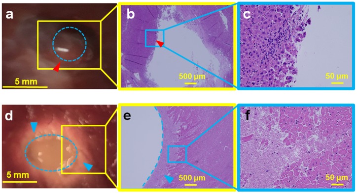
Fig 9. Ex vivo porcine liver (top panel) and cardiac muscle (bottom panel) sonicated at 600 W acoustic power at 1 Hz PRF and 20,000 cycles/pulse.
a. Gross pathology of the liver tissue with the lesion in the center (red arrowhead), showing minimal thermal damage with a liquefied central void (blue dotted circle). b. H&E slide showing the entire lesion with sharp boundaries (red arrowhead). c. Magnification (4X) of the Fig 9b, presenting intact cell structures at the periphery of the lesion. d. Gross pathology of the cardiac tissue with a large void in the center (blue dotted oval). A concentric ring of necrosis (blue arrowheads) surrounds the central void. e. H&E slide, with the void outlined by the blue dotted line and the blue arrowhead pointing to the region of necrosis. f. Magnified image (40X) of Fig 9e show regions of both necrosis and intact cellular structures.

