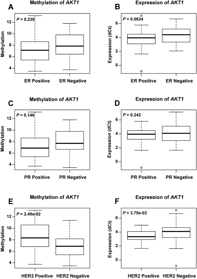
Fig 3. The methylation and expression level of AKT1 in breast cancer patients according to ER/PR/HER2 status.
Boxplots show the average methylation (A, C, E) and expression (B, D, F) level of AKT1 between ER-positive and ER-negative subgroups, between PR-positive and PR-negative subgroups, as well as between HER2-positive and HER2-negative subgroups. P values were calculated using the Wilcoxon signed rank test.

