FIGURE 1.
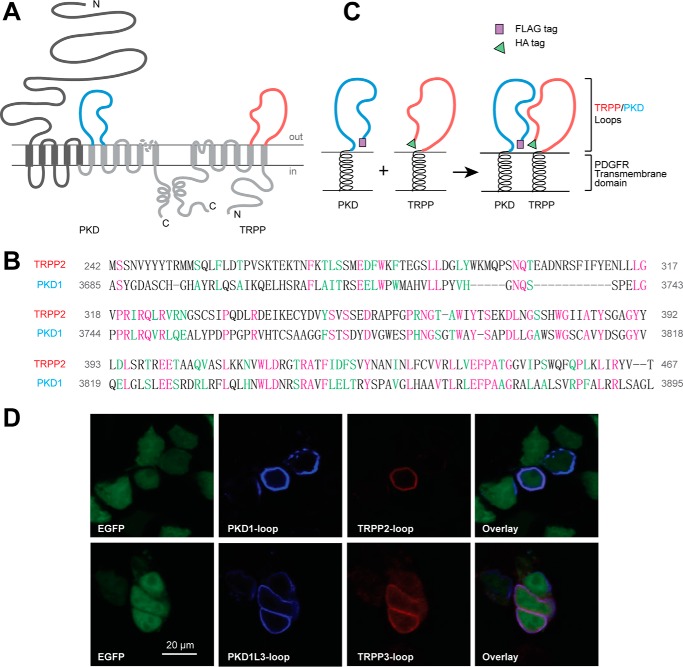
PKD and TRPP loop constructs, which were cloned into pDisplay vector and transfected into HEK 293T cells, expressed on the extracellular surface of the plasma membrane. A, the putative transmembrane topology of PKD (left) and TRPP (right) proteins, showing the position of extracellular loops between the sixth and seventh transmembrane domains of PKD proteins (S6-S7 loop, colored in blue) and between the first and second transmembrane domains of TRPP proteins (S1-S2 loop, colored in red). B, protein sequence alignment between the S1-S2 loop of human TRPP2 and the S6-S7 loop of human PKD1, two examples in the families. Purple, identical; green, conserved. Identity was 57 of 230 (25.0%), and similarity was 97 of 230 (42%). Amino acids positions in the full-length protein are labeled on both sides. C, schematic diagram of the structures of the expressed loop protein constructs whose association was confirmed in this study. cDNA of the loops were inserted into modified pDisplay vector (Life Technologies), which generated extracellular loop fragments with N-terminal FLAG or HA tag and C-terminal PDGFR transmembrane domain. D, surface co-localization of the FLAG-tagged PKD1 loop and HA-tagged TRPP2 loop and of the FLAG-tagged PKD1L3 loop and HA-tagged TRPP3 loop in HEK 293T cells. EGFP was co-transfected to label the cytosol. PKD loops and TRPP loops were stained first with anti-FLAG or anti-HA antibodies followed by Cy5 or Texas Red-labeled secondary antibodies on non-permeabilized cells.

