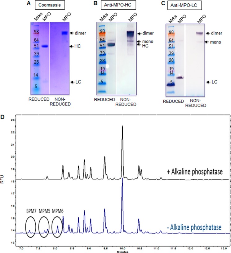
FIGURE 1.
Glycan analysis of MPO. A, SDS-PAGE analysis of MPO purified from human neutrophils under reducing and non-reducing conditions. B and C, Western blotting analysis of MPO as described in A using heavy chain MPO (B) and light chain MPO (C) antibodies. The bands corresponding to MPO monomer, dimer, mature HC and mature LC are indicated with arrows. D, oligosaccharide analysis of MPO by capillary zone electrophoresis. Phosphorylated peaks were digested to non-phosphorylated glycans with alkaline phosphatase (+AP). Indicated tentative peak IDs were assigned from co-migration with a known reference lysosomal enzyme standard included in the run. BPM7; bis-phosphorylated oligomannose 7; MPM5, monophosphorylated oligomannose 5; MPM6, monophosphorylated oligomannose 6. Results in A–C are representative of three separate experiments. Results in D are representative of two separate experiments. RFU, relative fluorescence units.

