FIGURE 4.
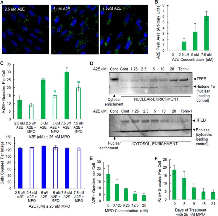
MPO promotes A2E clearance in a cell-based model of macular degeneration in the absence of cytotoxicity. A, representative high content images showing A2E autofluorescence in ARPE-19 cells preloaded with A2E for 6 h. B, detection of A2E parent molecule by UPLC in ARPE-19 cells preloaded with A2E for 6 h. C, quantification of high content images: cells preloaded with A2E, as in A, were incubated without (solid boxes) or with (open boxes) 25 nm MPO for 48 h. Top, A2E+ granules per cell: green. Bottom, nuclei number (cell number) per image: blue. D, Western blots of TFEB levels in nuclear and cytosol-enriched fractions from ARPE-19 cells preloaded with increasing concentrations of A2E as indicated or treated with 2 μm Torin-1. Blots were subsequently probed with nuclear and cytosolic marker proteins to confirm subcellular enrichment. Data are representative of three separate experiments. E, MPO dose-dependently promotes clearance of A2E in ARPE-19 cells. ARPE-19 cells were preloaded with 5 μm A2E for 6 h and then incubated with up to 25 nm MPO as indicated for 21 days. A2E autofluorescence was quantified by high content imaging. F, uptake of 25 nm MPO time-dependently promotes A2E clearance over 21 days in ARPE-19 cells preloaded with 5 μm A2E. Results in B, C, E, and F are expressed as the mean (n = 3 cultures) ± S.D. See “Experimental Procedures” for further details. Significant differences (p < 0.05) to control A2E groups.

