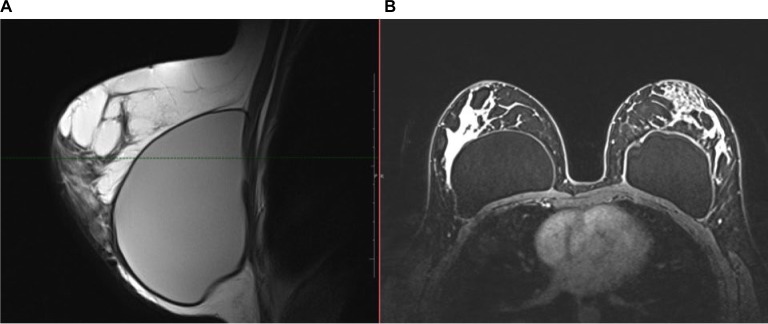
Figure 7.
A 43-year-old patient with thickening and discomfort in the left breast.
Notes: (A) Sagittal PD images of the left breast and (B) axial images after injection of the contrast agent show the increase in the anteroposterior diameter of the left breast implant, with thickening of the fibrous capsule and enhanced contrast compared with the right side.
Abbreviation: PD, proton density.

