
Figure 5. TβRII is deficient in inducing phosphorylation of TGF-β1/Smad pathway members and the expression of downstream target proteins.
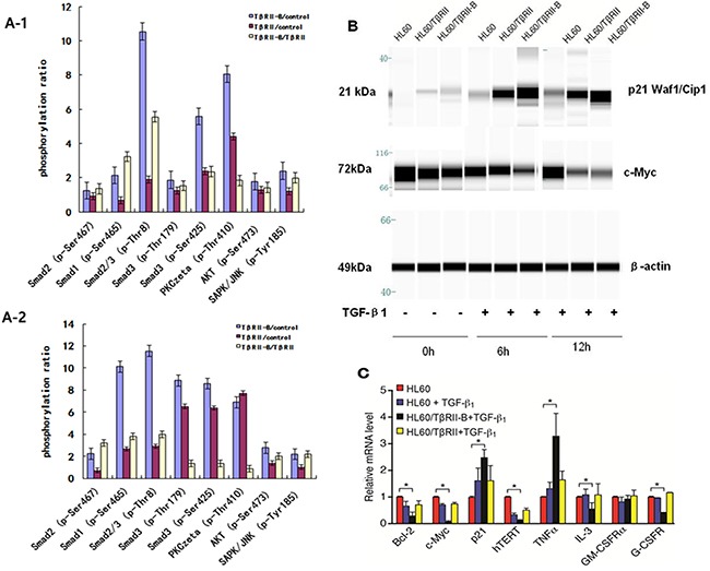
A1–2. Phosphorylation of TGF-β1/Smad pathway members increased significantly in HL60/TβRII-B cells compared to HL60/TβRII cells after incubation with 1 ng/mL TGF-β1 for 0.5 h or 2 h. B. Western blot analysis of c-Myc and p21CIP/WAF1 expression in TGF-β1-treated (1 ng/mL) HL60 cells that stably expressed different TβRII isoforms. Untreated HL60 cells served as a control. The data are representative of two independent experiments. C. Real-time PCR analysis of TGF-β1 targets in HL60 cells expressing either TβRII or TβRII-B. Untreated HL60 cells served as a control. The experiment was performed in triplicate. The data are expressed as the mean ± SEM. *P < 0.05, two-tailed Student's t-test.