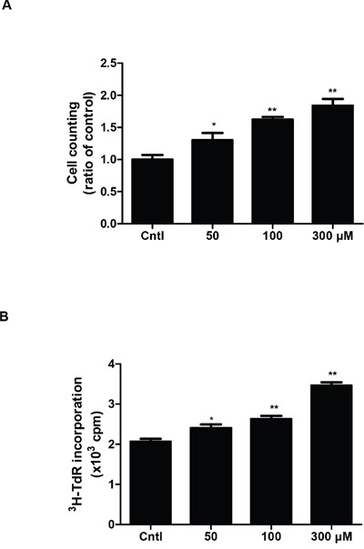
Figure 1. Effect of uric acid on cell proliferation in mouse mesangial cells.

A & B. After mesangial cells were cultivated to 60%–70% confluence, they were treated with uric acid at different doses (0, 50, 100, 300 μM) for 24 h and the cell proliferation was determined by cell counting (A) and [3H] thymidine (3H-TdR) incorporation (B). Values are means ± SE; n = 6 for each group. * P< 0.05 vs. control, ** P< 0.01 vs. control.
