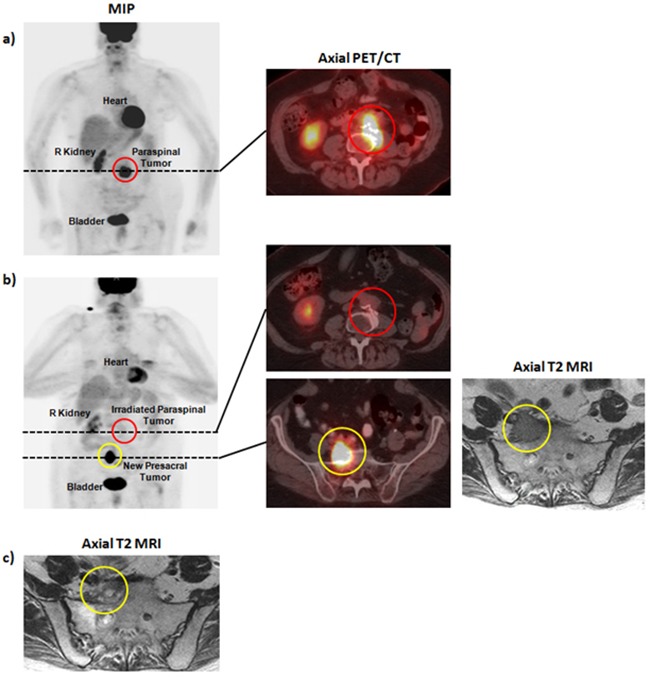
Figure 3. Pre- and post-therapy imaging for patient B.

a. Pre-radiotherapy PET/CT demonstrating a para-spinal mass involving the L4 vertebral body. b. Post-radiotherapy PET/CT and MRI demonstrating treatment response at L3-L4 and an out-of-field recurrence in a right pre-sacral lymph node. d. Post-radiotherapy MRI demonstrating reduction in size of the right pre-sacral lymph node.
