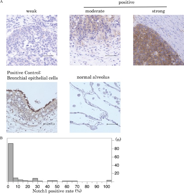
Figure 1. Notch1 expression in SCLC.

A. Immunohistochemical staining patterns of Notch1 are shown in SCLC. SCLC specimens were stained with anti-Notch1 antibody. Notch1 expression in normal bronchial and alveolar cells is also shown (scale bar = 50 μm). B. This histogram illustrates the distribution of Notch1 expression of any intensity in the 125 SCLC specimens that were investigated in the current study. The median Notch1-positive staining rate in SCLC tumors was 0% (95%CI:0-0.6). Cases with more than 5% staining were defined as the high Notch1 expression group.
