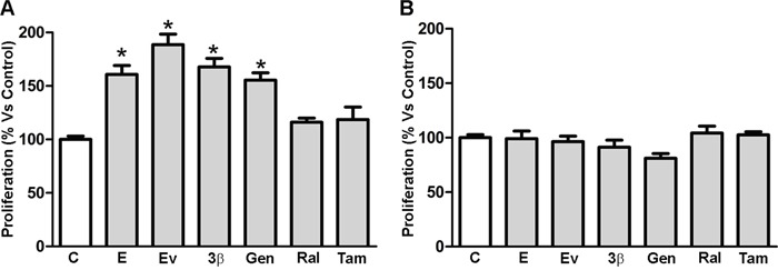
Figure 2. Cellular proliferation of MCF-7 and MDA-MB-231 cell lines. MCF-7.

A. and MDA-MB-231 B. cellular proliferation was evaluated by MTT assay 2 days after treatment with DMSO (Control), 17β-estradiol (10nM), estradiol valerate (10nM), 3β-Adiol (1μM), genistein (1μM), raloxifen (1μM) and tamoxifen (1μM). Statistical analysis was performed by one-way ANOVA followed by Bonferroni multiple comparison tests. *p<0.05 vs Control. Values represent the mean from three independent experiments. C. Control cells; E: 17β-estradiol; EV: estradiol valerate; 3β: 3β-Adiol; Gen: genistein; Ral: raloxifen; Tam: tamoxifen.
