
Figure 1. Baicalein prevents E2-induced cell growth, migration, and invasion in mammary epithelial cells.
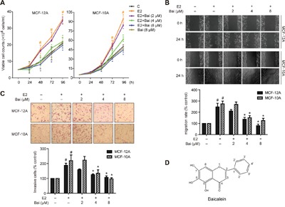
Cells were treated with E2 or E2 plus baicalein (Bai) for 5 weeks and were then used in the following experiments. A. Cell growth was measured using trypan blue exclusion assay. The cell growth curve represents the growth of cells in the different treatment groups over 4 days. B. Cell migration was measured using wound healing assay. Confluent monolayers were scratched and incubated in serum-free culture medium; images were captured at 0 and 24 h after wounding (magnification, ×100). The level of cell migration into the wound scratch was quantified as migration rate by comparing with the control (as 100%). C. Cell invasion was investigated using the Matrigel-coated transwell model. Invasive cells that passed through the membrane were evaluated using H&E staining (magnification, ×200). The results are expressed as invasive cells with respect to the control (as 100%). D. Chemical structure of baicalein. The images are representative of three independent experiments. Data are shown as means ± SEM (n = 3). *P < 0.05 vs. E2, #P < 0.05 vs. control.