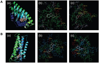
Figure 4. The binding of E2 and baicalein with ERα and GPR30.

A. Docking analyses for the binding of E2 and baicalein with ERα. (a) The three-dimensional model of E2 (orange) and baicalein (white) to ERα. (b) The binding of E2 to ERα is shown; hydrogen bonds with Glu 353, Leu 349, and Arg 394 sites were formed. (c) The binding of baicalein to ERα is shown, and hydrogen bonds with Glu 353, Leu 387, Leu346, and Arg 394 sites were formed. B. Docking analyses for the binding of E2 and baicalein to GPR30. (a) A three-dimensional model of the binding of E2 (orange) and baicalein (white) to GPR30. (b) The binding of E2 to GPR30 is shown, and hydrogen bonds with His 197, Asp 202, and Glu 218 sites were formed. (c) The binding of baicalein to GPR30 is shown, and hydrogen bonds with His 282, Asp 202, and His 200 sites were formed.
