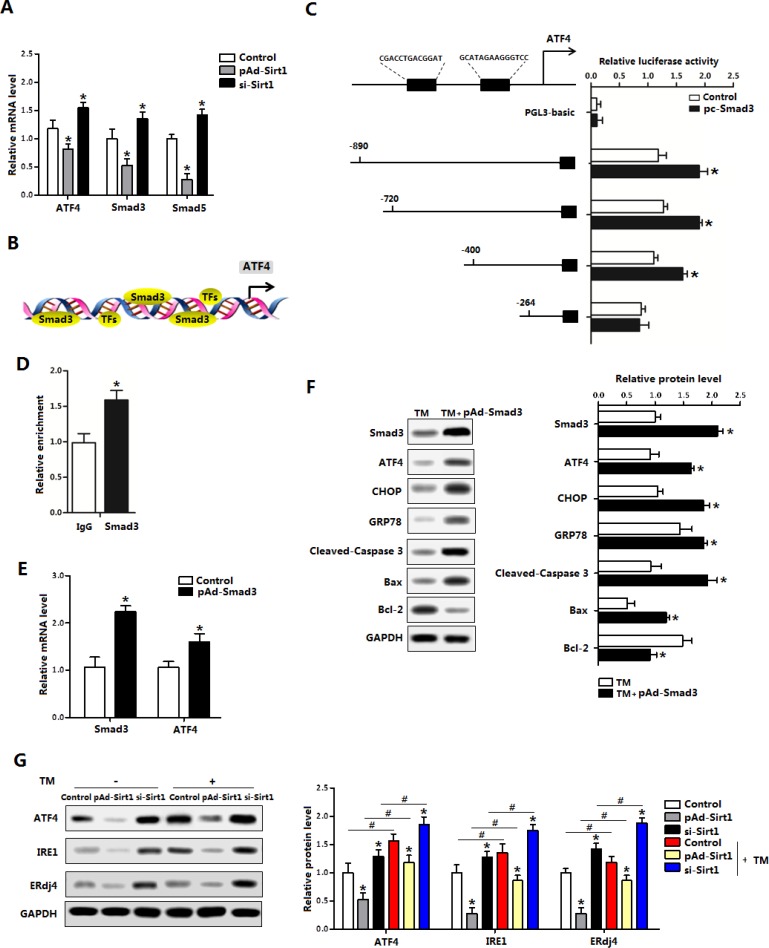
Figure 6. Smad3 promoted ATF4 transcription in brown adipocytes.
(A) Relative mRNA levels of ATF4, Smad3 and Smad5 of brown adipocytes pre-infected with pAd-Sirt1 or si-Sirt1 for 24 h (n = 3); (B) Predicate translation factors of ATF4 by using Genomatix software; (C) Dual luciferase reporter assay of ATF4 and Smad3. HEK293 cells were transfected with PGL3-basic or PGL3-ATF4 plasmids, and pc-Smad3 plasmid (n = 3); (D) ChIP analysis between ATF4 and Smad3 (n = 3); (E) Relative mRNA levels of Smad3 and ATF4 with pAd-Smad3 infection of brown adipocytes (n = 3); (F) Protein levels of Smad3, ATF4, CHOP, GRP78, Cleaved-Caspase3, Bax and Bcl-2 of brown adipocytes pre-incubated with TM and treated with pAd-Smad3 or not (n = 3); (G) Protein levels of ATF4, Smad3, IRE1 and ERdj4 of brown adipocytes pre-incubated with TM, and infected with pAd-Sirt1 or si-Sirt1 (n = 3). pAd-Smad3: recombinant adenovirus over-expression vector of Smad3, pc-Smad3: Overexpression plasmid of Smad3; pAd-Sirt1: recombinant adenovirus over-expression vector of Sirt1, si-Sirt1: recombinant lentiviral interference vector of Sirt1. Values are means ± SEM. *p < 0.05, #p < 0.05 compared with the control group.

