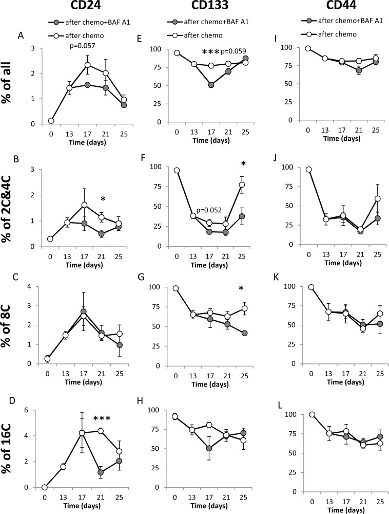
Figure 6. A single pulse of BAF A1 transiently affects stemness markers in senescent HCT116 cells.
(A–D) Evaluation of percentage of CD24 positive cells in cultures treated with the AFTER CHEMO or the AFTER CHEMO + BAF A1 protocol at various time points. Cells stained with H33342, as a ploidy discriminator, were probed with an anti-CD24-FITC antibody. Percentages of all cells (A), and CD24+ cells with 2C&4C (B), 8C (C) and 16C (D) DNA content were determined by flow cytometry. Cells labeled with isotypic IgGs were used as a negative control. (E–H) Evaluation of percentage of CD133+ cells at various time points. Cells stained with H33342 were probed with an anti-CD133-APC antibody. Percentages of all cells (E), and CD133+ cells with: 2C&4C (F), 8C (G) and 16C (H) DNA content were determined by flow cytometry. (I–L) Evaluation of percentages of CD44+ cells at various time points. Cells stained with H33342, were incubated with an anti-CD44-AlexaFluor700 antibody. Percentages of all cells (I), and CD44+ cells with: 2C&4C (J), 8C (K) and 16C (L) DNA content were determined by flow cytometry. Each bar represents mean ± SEM, N ≥ 3.*p < 0.05, **p < 0.01, ***p < 0.001 – AFTER CHEMO vs. AFTER CHEMO + BAF A1.

