
Figure 3. SAHA induces mast cell apoptosis, down-regulation of KIT and increase in H3K27ac.
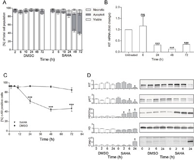
A. Effects of SAHA on cell death revealed a significant increase of apoptotic cell death at 48 h (p<0.01), as determined by flow cytometry by Annexin V and propidium iodide staining, n=3; B. A significant decrease of KIT mRNA was seen at 24 h (p<0.05, duplicates of 4 individual experiments), as was C. a decrease in the percentage of cell surface KIT positive cells (p<0.01 at 24 h,48 h and 72 h compared to baseline as well as to DMSO treated cells (triplicate of two separate experiments); D. Western blot analyses showed early responses to SAHA with increased H3K27ac already at 2 h, and decrease in phosphorylated KIT at 6 h, whereas total KIT decreased and active caspase 3 increased significantly at 24 h. Total histone H3 remained unchanged by SAHA treatment. *=p<0.05, **=p<0.01.